Figure 4.
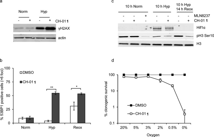
CH-01 induces DNA damage and is toxic in hypoxic conditions. (a) RKO cells were exposed to normoxia or hypoxia (≤0.02% O2) for 6 h in the presence or absence of 25 μM CH-01 (1). Western blots for γH2AX and actin are shown. (b) RKO cells were exposed to normoxia or hypoxia (≤0.02% O2) for 6 h and hypoxia followed by 18 h of reoxygenation ± 1 (25 μM). The graph shows the percentage of cells with >6 nuclear 53BP1 foci. Significance values: * p < 0.05; **p < 0.0001. (c) RKO cells were exposed to hypoxia for the time periods indicated with either 1 (25 μM), MLN8237 (500 nM) or, as a control, DMSO. The levels of phosphorylated histone 3 (pH3 Ser10) were determined by Western blotting. Histone 3 (H3) is shown as loading control. (d) Clonogenic assays were carried out on RKO cells exposed to the oxygen tensions indicated for 24 h in the presence of 25 μM 1 or DMSO.

