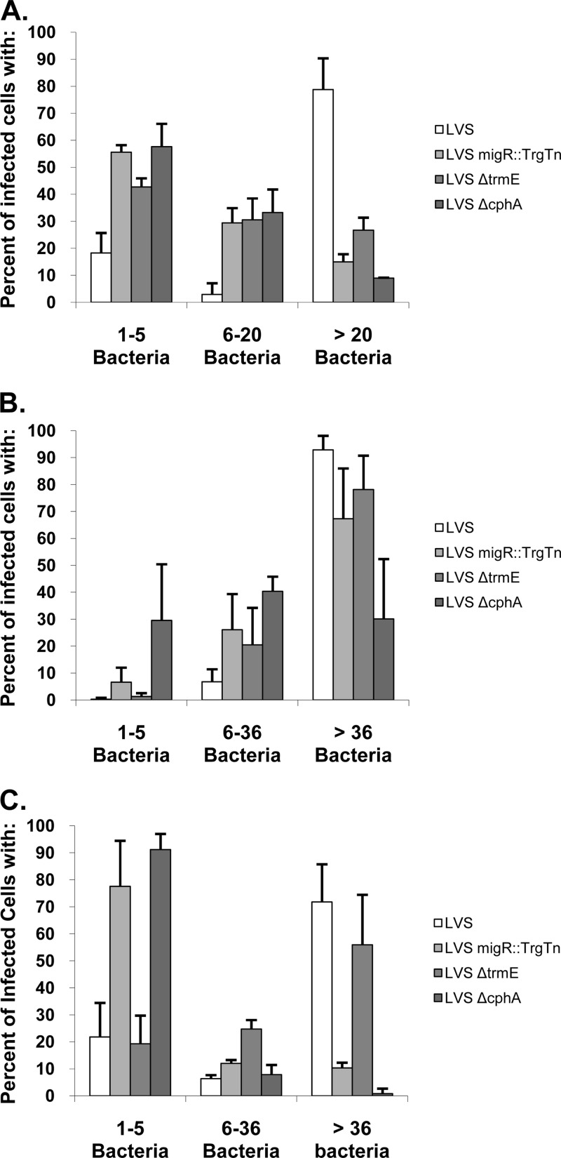
Fig 5.
Quantitative analysis of the cellular growth of F. tularensis mutants demonstrates that each strain has a unique pattern. MDMs, HEp-2 cells, and A549 cells were infected with LVS, LVS migR::TrgTn, LVS ΔtrmE, and LVS ΔcphA using the standard intracellular growth assay. At 24 h of growth, cells were fixed and the samples were prepared for confocal microscopy. The number of bacteria per infected cell was determined for each of the three cell lines via confocal microscopy, and the data are presented in the graphs. Greater than 100 infected cells were counted per strain and cell type. (A) MDMs; (B) HEp-2 cells; (C) A549 cells.

