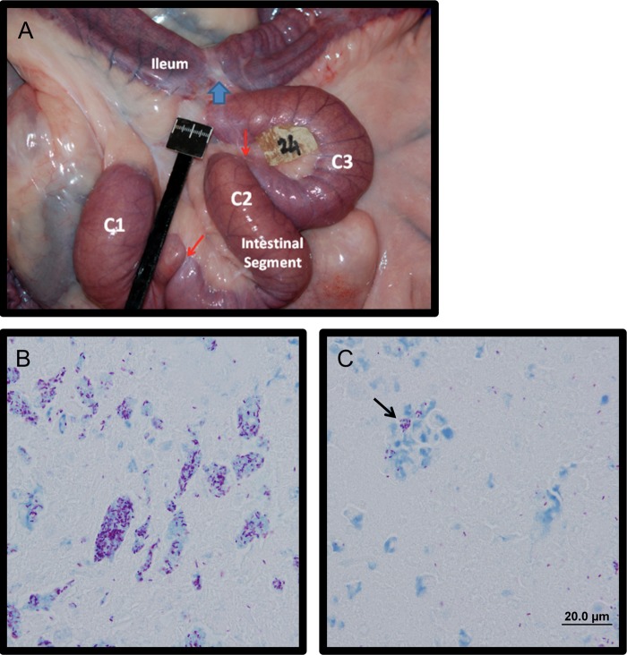
Fig 1.
Bovine calf intestines at 1 month after in vivo M. avium subsp. paratuberculosis infection. (A) Gross appearance of a surgically isolated distal intestinal segment in situ at 1 month postinfection. The surgically isolated segment was subdivided into three compartments, C1, C2, and C3, using silk ligatures (indicated by red arrows). The site where intestine proximal and distal to the isolated segment was anastomosed together is indicated with a blue arrow. (B) Ziehl-Neelsen stain of intestinal contents at 1 month after infection, showing diffuse aggregates of acid-fast Mycobacterium avium subsp. paratuberculosis at a magnification of ×100. (C)Acid fast-bacteria observed within cell remnants within the intestinal contents of an infected compartment (black arrow).

