
Table 2.
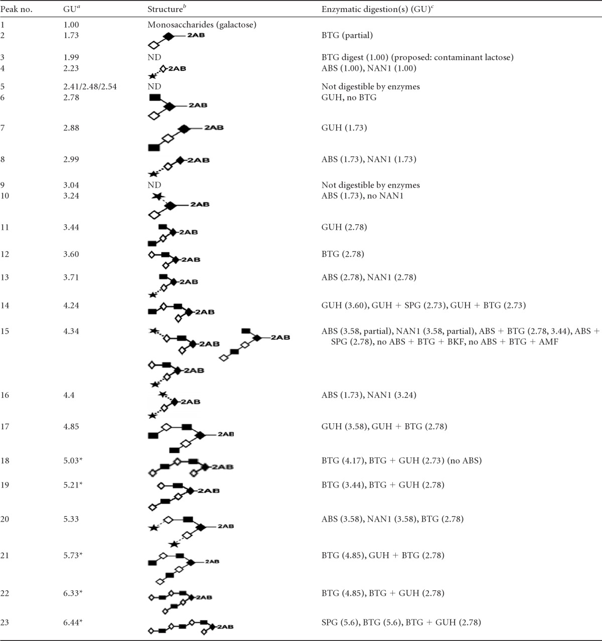
Structures of O-glycans released from E-12 mucins, described by HILIC-HPLC and exoglycosidase digestion arrays
a
Glucose unit (GU) values are averages of triplicates, with a standard deviation of 0.06. Asterisks indicate proposed isomeric structures.
b
Structures are represented using the Oxford system for monosaccharides and linkages (Fig. 6, key) (59). ND, not determined.
c
The enzymes used were bovine testis β-galactosidase (BTG), Arthrobacter ureafaciens sialidase (ABS), Streptococcus pneumoniae sialidase (NAN1), Streptococcus pneumoniae β-N-acetylglucosaminidase (GUH), Streptococcus pneumoniae β-galactosidase (SPG), bovine kidney α-fucosidase (BKF), and almond meal α-fucosidase (AMF).