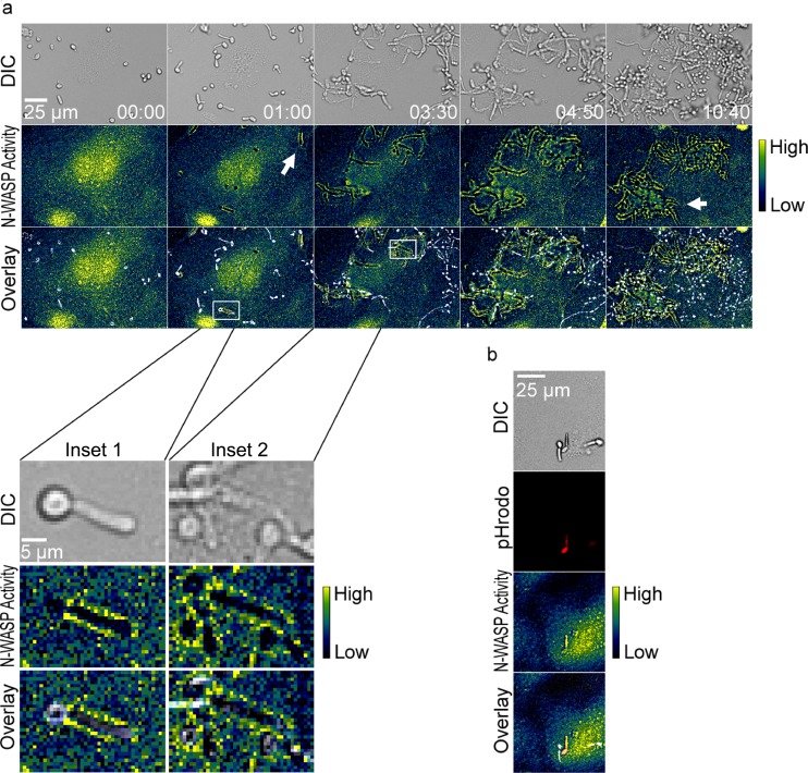
Fig 8.
(a) N-WASP activity also occurred at sites of internalization of live C. albicans SC5314. The top row shows DIC images, the middle row shows FRET (pseudocolor), and the bottom row shows overlays. A scale bar showing relative activity of N-WASP is shown on the right. White arrows show regions of N-WASP activity around internalizing yeast. Data are also shown in Movie S5 in the supplemental material. Inset 1 shows an enlargement from the 1-h time point, where N-WASP activity is observed around the germ tube but not around the mother cell. Inset 2 shows a later time point, when N-WASP activity is observed around both the germ tube and mother cell. Image sequences are representative of 3 independent experiments. (b) N-WASP activity occurs for internalization of pHrodo-labeled, heat-killed C. albicans hyphae. Here, N-WASP activity is observed around the internalized pHrodo-bright yeast but not for the adjacent pHrodo-dim yeast. Images are representative of 5 independent experiments.

