Fig 1.
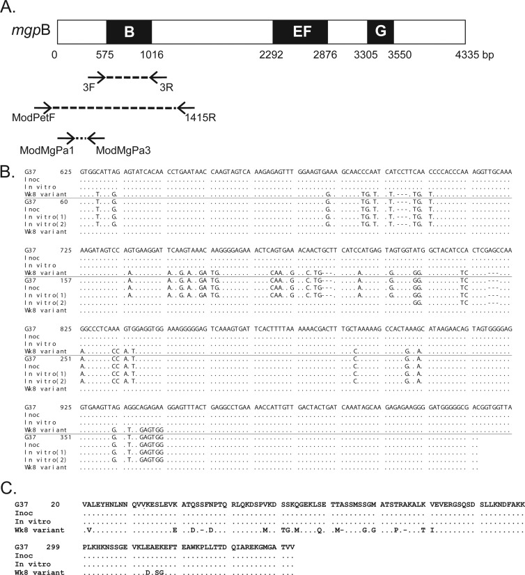
(A) Schematic of the mgpB expression site of M. genitalium G37-C. Black boxes (B, EF, and G) indicate the variable regions of mgpB, and intervening white areas represent conserved sequences (82). Arrows connected by broken lines show the location and direction of the primers used for PCR amplification. (B) Alignment of mgpB region B and MgPar8 DNA sequences obtained from the primate inoculum (Inoc), bacteria passaged in broth for 8 weeks (In vitro), and the week 8 primate specimen (Wk8 variant) with the published M. genitalium G37 type sequences. In each set of sequences, the top four lines represent mgpB expression site sequences, while the bottom five lines represent MgPar8 sequences. Nucleotides that are identical to the nucleotides in strain G37 are indicated by dots; nucleotides that differ from the G37 type sequence are shown. Deleted nucleotides are indicated by hyphens. Two MgPar8 sequences were identified in the in vitro-propagated control and are indicated as “In vitro (1)” and “In vitro (2)” (see Results). Nucleotides are numbered relative to the first base pair of G37 mgpB or MgPar8. (C) Alignment of the predicted MgpB region B amino acid sequences showing that the primate week 8 DNA sequence predicts an in-frame protein product with variant amino acids indicated by their single-letter abbreviation. Amino acids are numbered according to the start codon of MgpB.

