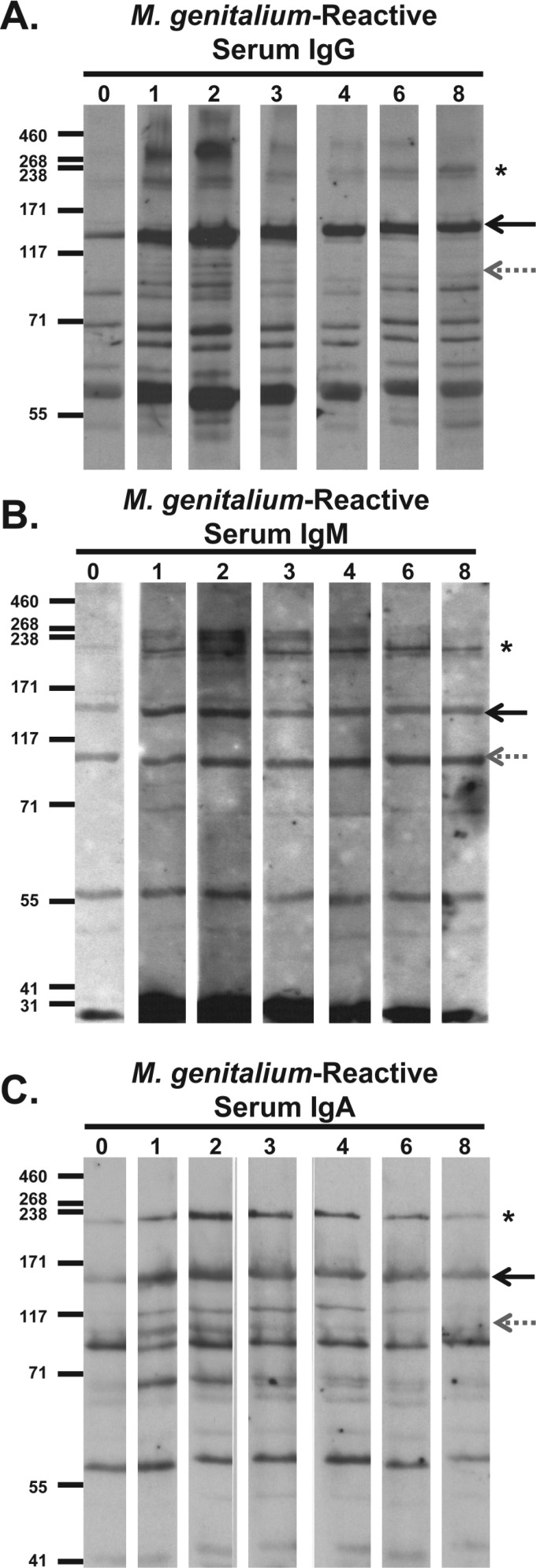
Fig 2.
Immunoblots of primate serum antibody reactivity to M. genitalium whole-cell lysates. Bacterial lysates were electrophoresed through an 8% SDS-polyacrylamide gel, transferred to membranes, and then cut into strips for reaction with primate serum samples collected at weeks 0 to 8 after infection. (A) Serum IgG reactivity. Primate serum samples were diluted 1:5,000, reac-tivity was detected using horseradish peroxidase (HRP)-conjugated goat anti-human IgG secondary antibody diluted 1:10,000, and the membranes were exposed to film for 6 min. (B) Serum IgM reactivity. Primate serum samples were diluted 1:800, reactivity was detected using HRP-conjugated goat anti-human IgM secondary antibody diluted 1:10,000, and then the membranes were exposed to film for 4 min. (C) Serum IgA reactivity. Primate serum samples were diluted 1:200, reactivity was detected using HRP-conjugated goat anti-human IgA secondary antibody diluted 1:8,000, and then the membranes were exposed to film for 13 min. The positions of MgpB (solid black arrows), MgpC (gray dotted arrows), and HMW2 (asterisks) are indicated to the right of the immunoblots. The positions of molecular mass markers (in kDa) are shown to the left of each immunoblot.

