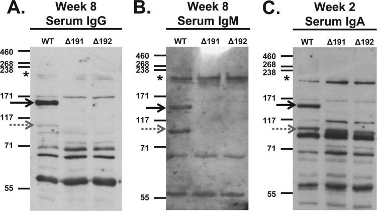
Fig 3.
Reactivity of primate serum antibodies to whole-cell lysates of wild-type M. genitalium G37-C (WT) and to mgpB (Δ191) and mgpC (Δ192) deletion mutants. (A) Serum IgG reactivity. Primate serum samples were diluted 1:5,000, reactivity was detected using HRP-conjugated goat anti-human IgG secondary antibody diluted 1:10,000 and then exposed to film for 5 min. (B) Serum IgM reactivity. Primate serum samples were diluted 1:800, and reactivity was detected using HRP-conjugated goat anti-human IgM secondary antibody diluted 1:10,000 and then exposed to film for 4 min. (C) Serum IgA reactivity. Due to the limited volume of week 8 serum samples available, the reactivity of week 2 IgA is shown. Primate serum samples were diluted 1:200, and reactivity was detected using HRP-conjugated goat anti-human IgA secondary antibody diluted 1:8,000 and then exposed to film for 20 min. The positions of MgpB (solid black arrows), MgpC (gray dotted arrows), and HMW2 (asterisks) are indicated. The positions of molecular mass markers (in kDa) are shown to the left of each immunoblot.

