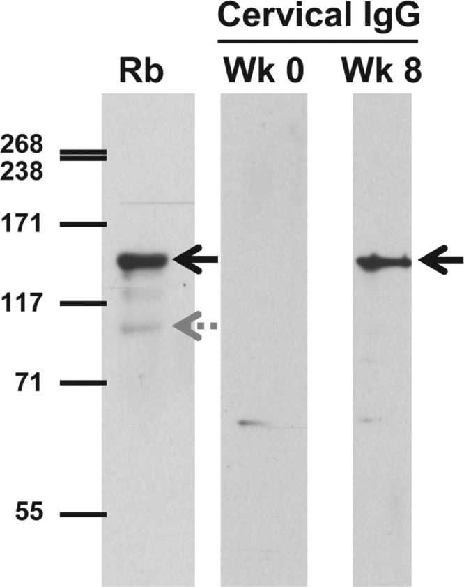
Fig 4.

Immunoblot analysis of primate genital tract antibody reactivity to whole-cell lysates of wild-type M. genitalium. Cervical exudates collected prior to inoculation (week 0 [Wk 0]) or after 8 weeks of infection (Wk 8) were diluted 1:50 and reacted with whole-cell lysates of M. genitalium G37-C. To identify the positions of the 140-kDa MgpB and 110-kDa MgpC proteins, bacterial lysates were reacted with antibodies produced in rabbits (Rb), combined, and diluted 1:10,000. HRP-conjugated secondary antibody (anti-human IgG or anti-rabbit IgG) was diluted 1:10,000. After reaction with chemiluminescent reagents, membranes were exposed to film for 5 min or 1 min, respectively. The positions of MgpB (solid black arrows) and MgpC (gray dotted arrow) are indicated.
