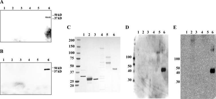
Fig 1.
Western immunoblot assays of patient serum to detect levels of IgG (A) and IgM (B) antibodies to various pathogens. Lanes: 1, L. interrogans (recombinant LipL32 and LipL41 proteins); 2, Rickettsia conorii (recombinant OmpA protein fragments); 3, C. burnetii (recombinant Com-1 protein); 4, R. typhi (whole-cell antigen from R. typhi strain Wilmington); 5, R. typhi (recombinant OmpB protein fragments); 6, O. tsutsugamushi (recombinant R56 proteins from O. tsutsugamushi strains Karp, Kato, Gilliam, and TA763). The recombinant antigens/whole-cell antigens derived from various pathogens were separated on a 4 to 15% polyacrylamide gel, transferred to a nylon membrane, and probed with patient serum (1:100 dilution) overnight at 4°C. The blot was probed with a horseradish peroxidase-conjugated anti-human IgG or IgM secondary antibody (1:1,000 dilution) and visualized with a chemiluminescence-based detection kit (Amersham). (C) Corresponding protein gel stained with SimplyBlue SafeStain (Invitrogen) showing the presence of recombinant/whole-cell antigens used in a Western blot assay loaded in the same order as in panels A and B. (D and E) Positive-control Western blot assays detecting the antigens (loaded in the same order as in panels A and B) in the serum of a confirmed scrub typhus patient. The patient's scrub typhus was confirmed by a 4-fold titer increase and isolation of O. tsutsugamushi from a serum sample. Panel D shows IgG results, and panel E shows IgM results. The values to the left of panels C to E are molecular sizes in kilodaltons.

