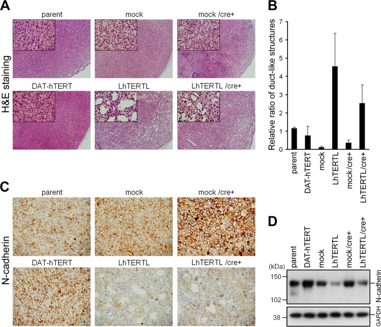
Fig 3.
PC-3 cell differentiation due to telomere elongation. (A) H&E-stained tissue sections. PC-3/DAT-hTERT cells (lower left panel) remained in an undifferentiated state similar to that for control cells (upper panel), whereas PC-3/LhTERTL and PC-3/LhTERTL/cre+ cells showed duct-like structures (lower center and right panels). Original magnification, ×40. Insets, magnified views (×200). (B) Quantification of duct-like structures. Parameters are the average and standard deviation of the number of ducts per unit area in each tumor (n = 2 or 3). Reproducibility and significant differences were assessed using additional xenograft tumor samples for microarray experiments (see Fig. S3 in the supplemental material). (C) Immunohistochemistry of N-cadherin. Suppression of N-cadherin expression was observed in PC-3/LhTERTL and PC-3/LhTERTL/cre+ cells (lower center and right panels). Original magnification, ×200. (D) Western analysis demonstrated that the reduction of N-cadherin expression in telomere-elongated PC-3 cells before subcutaneous injection.

