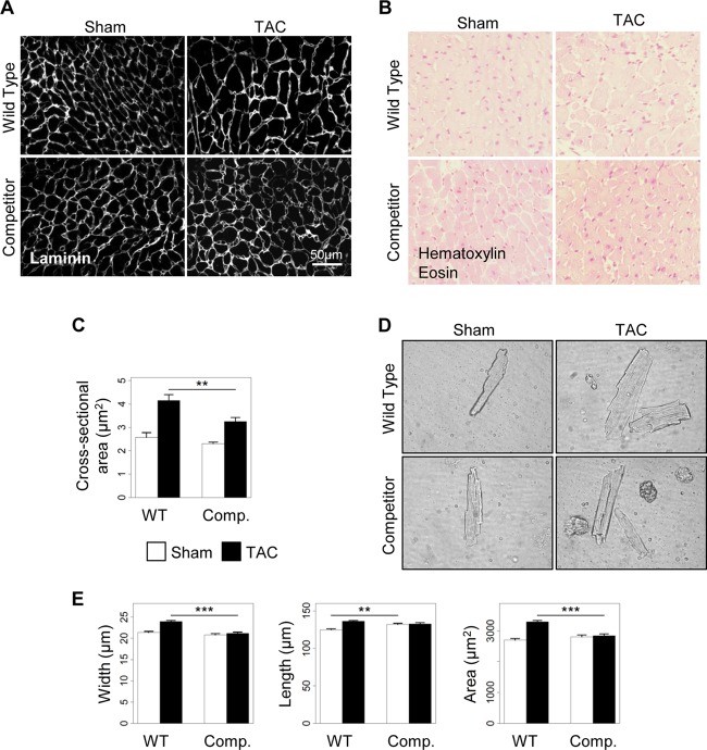
Fig 8.
Disruption of the AKAP-Lbc/p38 signaling complex inhibits cardiomyocyte hypertrophy in response to pressure overload. Transgenic mice overexpressing the competitor fragment of AKAP-Lbc (Comp.) and their WT littermates were subjected to 2 weeks of TAC or a sham operation. (A and B) Antilaminin (A) and hematoxylin and eosin (B) stainings of transversal heart sections from the different groups of mice. (C) Quantification of cardiomyocyte cross-sectional areas from the indicated groups (WT-sham, n = 6; WT-TAC, n = 7; competitor-sham, n = 5; competitor-TAC, n = 9; an average of 350 cells were analyzed per mouse). (D) Representative cardiomyocytes (phase-contrast images) isolated from WT and transgenic (Comp.) animals subjected to 2 weeks of aortic constriction. (E) Quantitative analysis of the width, length, and area (width times length) of cardiomyocytes from hearts of the indicated groups of mice (WT-sham, n = 3; WT-TAC, n = 6; competitor-sham, n = 3; competitor-TAC, n = 3; an average of 500 cells were analyzed per mouse). Values are presented as means and SEM. Statistical differences were analyzed using the Student t test. **, P < 0.01 versus WT-TAC; ***, P < 0.0001 versus WT-TAC.

