
Fig 9.
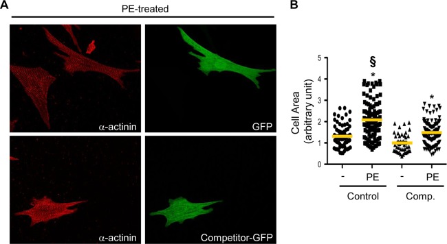
The AKAP-Lbc competitor fragment impairs the hypertrophic response of rat NVMs induced by α1-AR stimulation. (A) Rat NVMs were transfected with GFP (control) or with the GFP-tagged AKAP-Lbc competitor fragment (Competitor-GFP). Seventy-two hours after transfection, cardiomyocytes were serum starved for 24 h and incubated for an additional 24 h in the absence or presence of 10−4 M PE. The cells were then fixed, permeabilized, and incubated with anti-α-actinin (red) monoclonal antibodies, as well as rhodamine-conjugated anti-mouse secondary antibodies. GFP expression was visualized directly by fluorescent excitation at 490 nm. (B) Mean cell surface areas of cardiomyocytes transfected and treated or not with PE. The cell surface area was determined on a total of 120 GFP-positive cardiomyocytes derived from four independent experiments by using Image J software. Statistical significance was analyzed using an ANOVA test followed by Tukey posttests with Bonferroni corrections. *, P < 0.05 compared with the cell surface area measured in nonstimulated cardiomyocytes transfected with GFP (Control). §, P < 0.05 compared with the cell surface area measured in PE-stimulated cardiomyocytes transfected with GFP (Control).