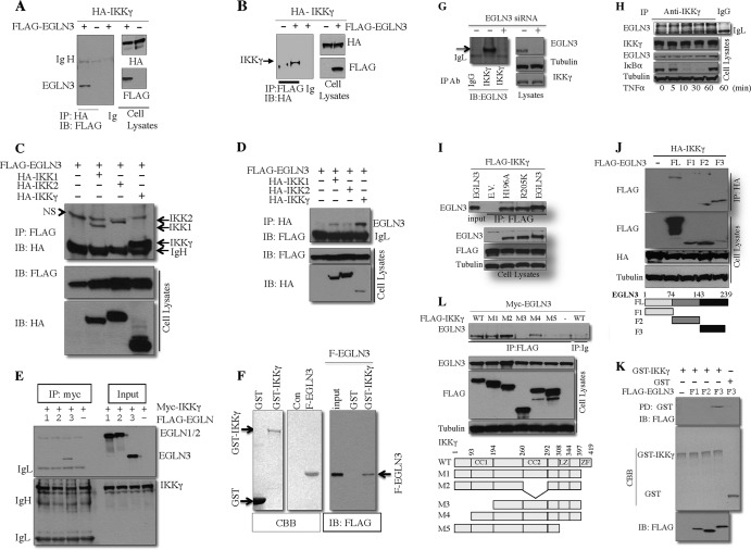
Fig 2.
EGLN3 interacts with IKKγ. (A to E) Total cell lysates from HEK293T transfected with the indicated plasmids were immunoprecipitated with anti-HA (A and D), anti-FLAG (B and C), anti-myc (E), or mouse IgG. The immune complex and cell lysates were analyzed by immunoblotting (IB). (F and K) Purified FLAG-EGLN3 or its fragments (F1, F2, or F3) were incubated with GST alone or GST-IKKγ immobilized on glutathione-Sepharose beads. The precipitates or purified FLAG-EGLN3 fragments were analyzed by immunoblotting. Purified FLAG-EGLN3, GST, and GST-IKKγ were visualized by Coomassie blue staining. Con, control; NS, nonspecific band. (G and H) Total cell lysates from HeLa cells treated with the EGLN3 siRNA (or control siRNA) (G) or from HeLa cells treated with TNF-α (10 ng/ml) (H) were immunoprecipitated with anti-IKKγ or control IgG. The immune complex and cell lysates were analyzed by immunoblotting. (I, J, and L) HEK293T cells were transfected with the indicated plasmids. Cell lysates were immunoprecipitated with anti-FLAG (I and L) or anti-HA (J). The immune complex and cell lysates were examined by immunoblotting. The bottom of panel J shows the schematic of wild-type EGLN3 and its fragments used; the bottom of panel L shows a schematic representation of wild-type (WT) IKKγ and its mutants used. FL, full-length EGLN3; CC1, coiled-coil domain 1; CC2, coiled-coil domain 2; LZ, leucine zipper region; ZF, zinc finger region; IP, immunoprecipitation; IgH, heavy chain of IgG; IgL, light chain of IgG; Ig, immunoglobulin; EV, empty vector; CBB, Coomassie blue staining; F-EGLN3, FLAG-tagged EGLN3.

