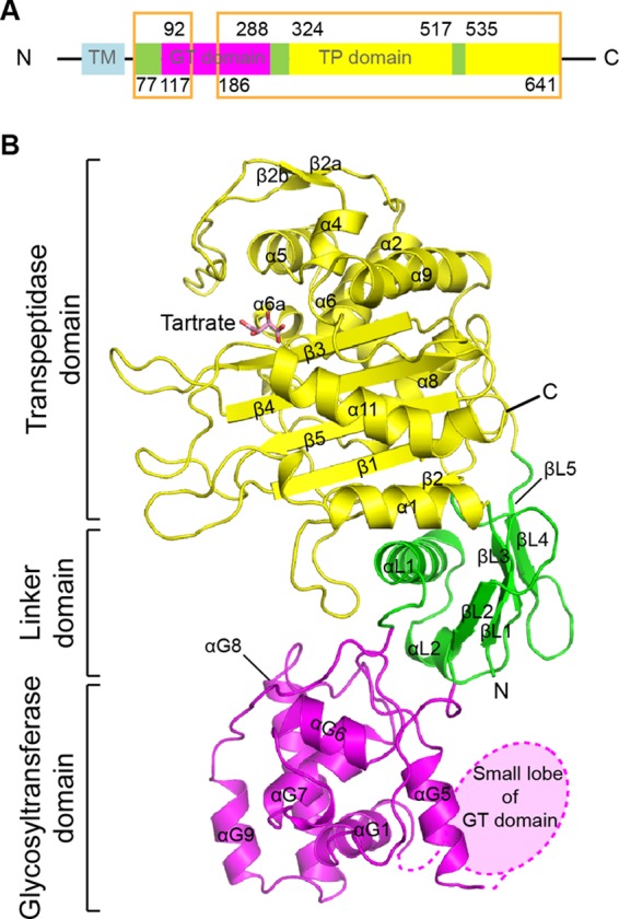
Fig 1.

Structure of PBP4 from L. monocytogenes. (A) Schematic representation of the organization of LmPBP4 domains. The transmembrane helix (TM), GT, linker, and TP domains of LmPBP4 are sky blue, magenta, yellow, and green, respectively. The regions presented in the crystal structures are boxed. (B) Ribbon representation of the apo-LmPBP4 structure. The secondary structural elements are colored as in panel A. A tartrate molecule bound to the active site is presented as a stick model. In this orientation, the TP domain is at the top, and the GT domain is at the bottom.
