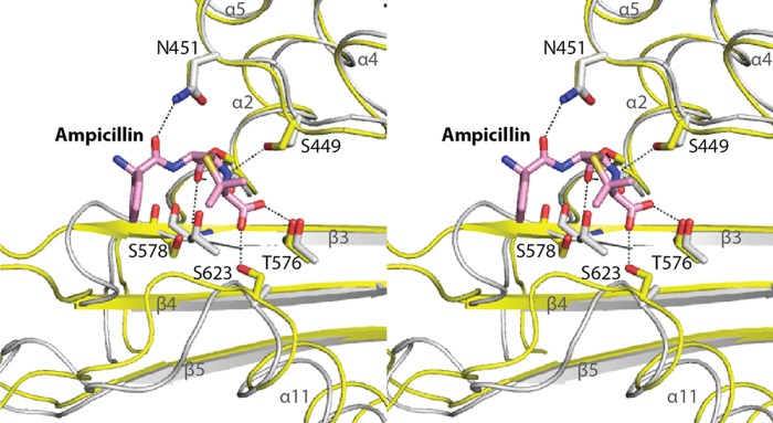
Fig 4.

Comparison of β-lactam recognition between LmPBP4 and EcPBP1b (PDB code 3FWM). The two structures are superimposed and shown in stereoview. Atoms of LmPBP4 are colored as described in the legend to Fig. 1. EcPBP1b is colored gray, and the secondary structure is presented as a ribbon model. The bound ampicillin is shown as a stick model.
