Fig 5.
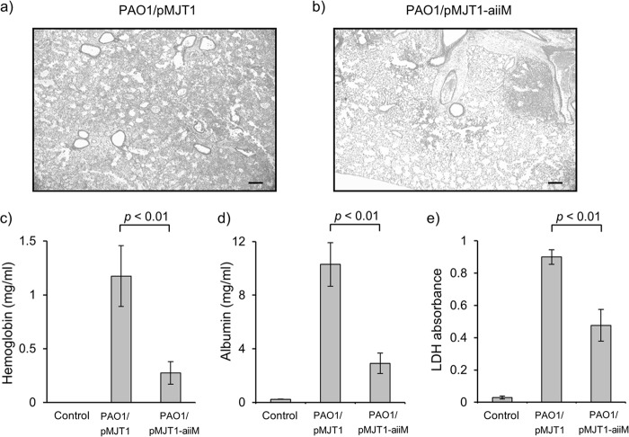
Lung tissue damage caused by P. aeruginosa infection. Histological examination of the lungs from the mice infected with P. aeruginosa PAO1/pMJT1 (a) and PAO1/pMJT1-aiiM (b). Lung tissue sections were obtained at 24 h postinfection and stained with hematoxylin and eosin. Representative images are shown at an original magnification of ×25. Scale bar, 200 μm. The amount of hemoglobin (c), albumin (d), and LDH (e) in cell-free BAL fluids at 24 h after infection with P. aeruginosa PAO1/pMJT1, PAO1/pMJT1-aiiM, or uninfected control is also shown. Each bar represents average values, and error bars show standard errors of the means (n = 8 mice per group). Mice infected with P. aeruginosa PAO1/pMJT1-aiiM developed only small lung lesions and had significantly decreased levels of hemoglobin, albumin, and LDH in BAL fluids.

