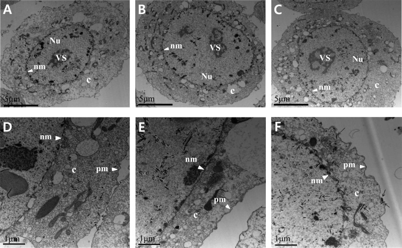
Fig 7.
Transmission electron microscopy analysis of Sf9 cells transfected with Ac-MK (A and D), Ac78KO (B and E), or Ac78Re (C and F) at 24 h p.t. (A, B, and C) Enlarged nucleus (Nu) and virogenic stroma (VS) in Ac-MK-, Ac78KO-, or Ac78Re-transfected cells. c, cytoplasm; nm, nuclear membrane. (D and F) Higher-magnification micrographs of Ac-MK- or Ac78Re-transfected cells displaying normal nucleocapsids residing in the cytoplasm (c) and budding from the plasma membrane (pm). (E) In Ac78KO-transfected cells, nucleocapsids and masses of electron-lucent tubular structures (arrows) were observed in the nucleus, but no nucleocapsids were observed in the cytoplasm.

