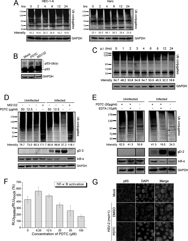
Fig 6.
PDTC may inhibit HSV replication through dysregulation of the UPS and inhibition of NF-κB activation. (A) HSV-2 infection changed the cellular profile of ubiquitin conjugates. Cells were incubated with 50 μg/ml PDTC and collected at the indicated time points. The levels of ubiquitin conjugates were determined via Western blot assay. (B) PDTC induced the accumulation of ubiquitinated p53. HEC-1-A cells were cultured in the presence of PDTC (50 μg/ml) or MG132 (6 μg/ml) for 2 h. p53 and its ubiquitinated forms were visualized using p53-specific antibody via Western blot assay. (C) HSV-2 infection changed the cellular profile of ubiquitin conjugates. HEC-1-A cells infected with HSV-2 (MOI = 1) were collected at the indicated times, and the levels of ubiquitin conjugates were determined. (D) PDTC inhibited HSV replication through dysregulation of the UPS and inhibition of HSV-induced IκB-α degradation. Cells treated with PDTC or MG132 were either mock infected or infected with HSV-2 (MOI = 1). The levels of ubiquitin conjugates, gD, and IκB-α were determined 24 h p.i. (E) PDTC-induced dysregulation of the UPS could be mitigated by EDTA. Cells exposed to PDTC were either mock treated or treated with EDTA (10 μM). The levels of ubiquitin conjugates, gD, and IκB-α were determined 24 h p.i. (F) PDTC treatment inhibited HSV-2-induced NF-κB activation. NF-κB activation was determined using NF-κB-luc reporter plasmid as described in the text. The RLU were determined 24 h p.i. and are expressed as the percentage of the amount in the mock-infected cells. (G) PDTC disrupted HSV-2-induced p65 nuclear translocation. p65 translocation in uninfected or infected cells treated with dimethyl sulfoxide (DMSO) or PDTC (50 μg/ml) was determined by immunofluorescence staining 24 h p.i. as described in the text. All experiments were performed 2 or 3 times, and the results of a representative experiment are shown. Error bars show standard deviations.

