
Fig 2.
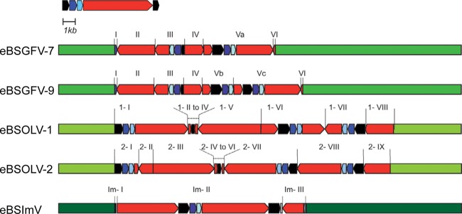
Overview of eBSV structures in PKW. Banana genomic sequences are in green. The BSV genome is represented in linear view with dark blue, light blue, and red boxes indicating ORF 1, ORF2, and ORF3 of the virus, respectively. The intergenic region is in black. eBSV fragments are indicated.