
Fig 9.
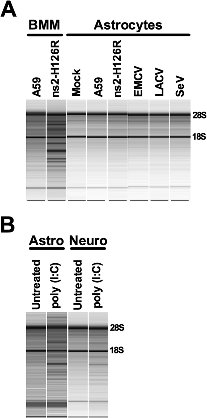
Activation of RNase L by other viruses and poly(I·C). (A) BMM and astrocytes were infected with A59, ns2-H126R, EMCV, LACV, and SeV (1 PFU/cell of SeV and LACV and 0.1 PFU/cell of EMCV). RNA was extracted from infected cells, and rRNA degradation was assessed with a Bioanalyzer. The positions of 28S and 18S rRNAs are indicated. The data shown are taken from three separate chips: (i) BMM RNA; (ii) SeV-infected cell RNA; and (iii) mock-, A59-, ns2-H126R-, EMCV-, and LACV-infected cell RNA. (B) Astrocyte and neuron cultures were treated with 100 U/ml IFN for 24 h and transfected with 10 μg/ml poly(I·C) with Lipofectamine 2000. Six hours later, RNA was extracted, and rRNA degradation was assessed with a Bioanalyzer. Astrocyte and neuron RNAs were analyzed on separate chips. The data are from one representative of two experiments.