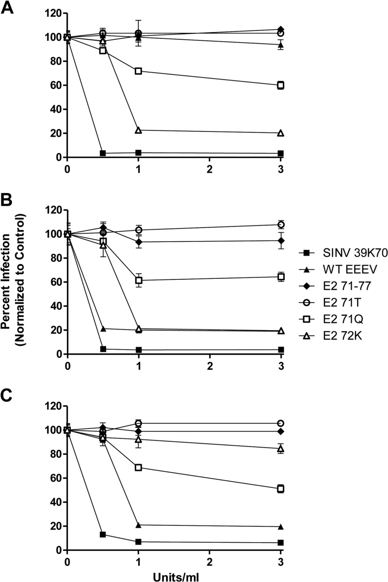
Fig 3.
Sensitivity of infection by natural variants to heparinase digestion. BHK cell monolayers were washed with VD and then digested with either heparinase I (A), heparinase II (B), or heparinase III (C) for 1 h at 37°C and washed 2 times with VD prior to infection with the indicated viruses. At 48 h postinfection, plaques on the monolayers were enumerated. Data are presented as changes in infectivity with increasing heparinase concentrations, normalized to a no-heparinase control set to 100% infectivity. Error bars are standard deviations, and some are too small to be seen.

