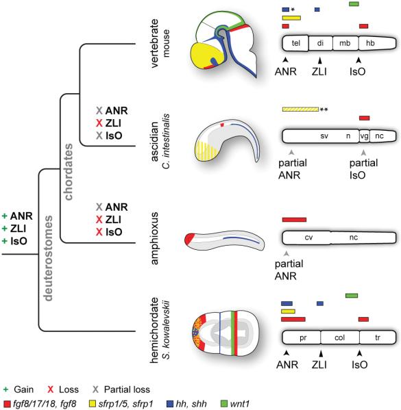
Figure 4. Evolutionary gain and loss of ANR, ZLI, and IsO-like genetic programs.
Schematic diagrams depicting expression of fgf8, sfrp1, shh, and wnt1 homologs in the mouse brain and ectoderm of C. intestinalis, amphioxus, and S. kowalevskii. Embryos are oriented with anterior to left and dorsal to top. Bar diagrams are oriented with anterior to the left. Diagrams depict only expression domains related to signalling components of vertebrate CNS signalling centres. Abbreviations: cv, cerebral vesicle; n, neck; nc, nerve cord; sv, sensory vesicle; vg, visceral ganglion; others as described previously. Diagrams not to scale. *Note that shh is expressed in the medial ganglionic eminence, adjacent to the ANR.**Sfrp1/5 is expressed in the C. intestinalis anterior ectoderm from 64-cells up to neurulation, but is then downregulated in the anterior ectoderm and CNS (depicted as yellow stripes).

