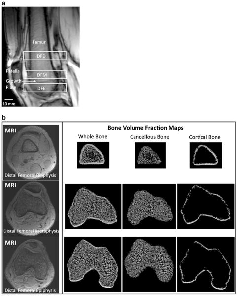
Fig. 1.
a Sagittal scout MR localizer image of the distal femur demonstrating the analysis locations for the 10-mm-thick volumes of interest (VOIs) at the distal femoral diaphysis (DFD), distal femoral metaphysis (DFM), and distal femoral epiphysis (DFE). These were defined anatomically by the superior pole of the patella (inferior margin of the DFD) and the healed growth plate (inferior margin of the DFM, superior margin of the DFE). b Representative axial MR images (left panel) at the distal femoral diaphysis (top row), distal femoral metaphysis (middle row), and distal femoral epiphysis (bottom row), and corresponding bone volume fraction maps of whole, cancellous, and cortical bone (right panel) from the same levels. In MR images of bone microarchitecture, trabeculae are represented by hypointense (dark) linear structures and marrow is hyperintense (white). For the bone volume fraction maps, MR images were inverted to create images with bone in white

