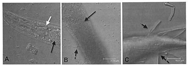
Fig. (1). Progression of Candida albicans infection in the C. elegans intestine.

(A). The nematode becomes infected with fungi by consuming the yeast cells as a food source. The white arrow points to the C. elegans pharyngeal grinder. Black arrows point to the location of the fungal cells in the nematode intestinal lumen. (B). Once the C. albicans cells are in the nematode, they form filaments (indicated by the broken black arrows) (C) that penetrate through the worm cuticle.
