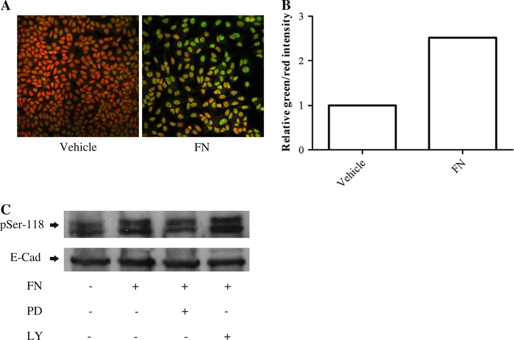
Fig. 7.
Fibronectin induces phosphorylation of ER at serine-118 in MCF-7 cells through MAPK/ERK1/2. a MCF-7 cells were grown to 80% confluency and starved for 48 h in phenol red free DMEM/F12. They were then treated with vehicle (water) or FN (30 µg/ml) for 15 min. Phospho-serine-118 ER was detected by immunofluorescence as described for LM05-E cells in Fig. 6. We found an increase in the levels of pSer-118 by treatment with FN. b Quantification of the staining intensity for pSer-118 corresponding to images shown in (a). Values for pSer-118 were normalized to propidium iodide for each treatment. c Western blot showing phosphorylation of ER at serine- 118 in MCF-7 cells. MCF-7 cells were grown to 80% confluency and then starved for 48 h. To determine the involvement of the MAPK/ ERK1/2 and PI3K/AKT pathways on the phosphorylation of ER-α, starved cells were pre-treated for 1 h with either vehicle (DMSO), PD98059 (10 µM), or LY294002 (10 µM). They were then subjected to a 15-min pulse of FN (30 µg/ml) or water as a control. Western blot analysis shows that FN leads to the phosphorylation of ER-α, and that it is inhibited by PD98059. No effect was detected with LY294002. One of at least two experiments is shown

