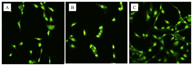
Fig. 7.

Confocal microscopy images of tissue cells (MC-3T3) grew on PET film (A), PET film treated using low power discharge oxygen (60 watts, 30 minutes) (B), and regenerated PET film from biofilm contaminated films using low power discharge oxygen (60 watts, 30 minutes) (C). Cells were cultured on PET films for two days and then stained using Live/Dead kit.
