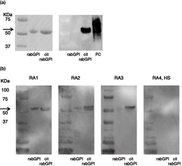
Figure 4.

(a) Immunoblotting of native rabbit glucose-6-phosphate isomerase (GPI) and citrullinated rabbit GPI (cit rabGPI). Bands from Gelcode Blue stain after sodium dodecyl sulphate-polyacrylamide gel electrophoresis (SDS-PAGE) were identified as rabGPI and cit rabGPI (left). Anti-modified citrulline antibodies demonstrated the presence of citrullinated protein at 60 kDa only in cit rabGPI well (right). Arrow shows 60 kDa. (b) Identification of anti-rabGPI and anti-cit rabGPI antibodies by Western blotting. The same reaction was shown to rabGPI and cit rabGPI in rheumatoid arthritis (RA) group 1 (RA1). However, a stronger reaction was shown to citrullinated GPI in RA2 and RA3. Neither of them was detected in RA4 and healthy control subjects (HS). Arrow shows 60 kDa.
