
Figure 2.
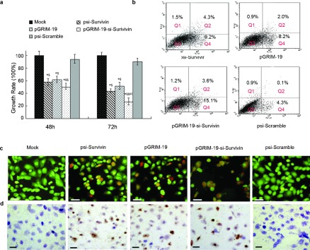
Growth inhibition and apoptosis assays in DU145 cells transfected with different treatment plasmids. (a) Growth inhibitory rate for DU145 cells transfected with psi-Survivin, pGRIM-19, pGRIM-19-si-Survivin and psi-Scramble (*P<0.05, **P<0.01 versus mock; §P<0.05, §§P<0.01 versus psi-Scramble group; #P<0.05 versus psi-Survivin; ‖‖P<0.05 versus pGRIM-19 groups). (b) Detection via FCS of apoptosis stimulated by recombinant treatment plasmids, Q1=dead cells, Q2=late apoptotic cells, Q3=normal cells, Q=early apoptotic cells. (c) Representative fluoromicrographs of apoptosis detected by AO/EB assay. The orange labeling indicates early stage apoptotic cells and the red labeling indicates late stage apoptotic cells. (d) Apoptosis of DU145 cells treated with psi-Survivin, pGRIM-19, pGRIM-19-si-Survivin and psi-Scramble plasmids by TUNEL staining, the brown labeling indicates the apoptotic cells (scale bar=20 µm). AO/EB, acridine orange/ethidium bromide; FCS, fetal calf serum; GRIM-19, gene associated with retinoid-interferon-induced mortality-19; RT-PCR, reverse transcription PCR; TUNEL, terminal deoxynucleotidyl transferase-mediated nick end labeling.