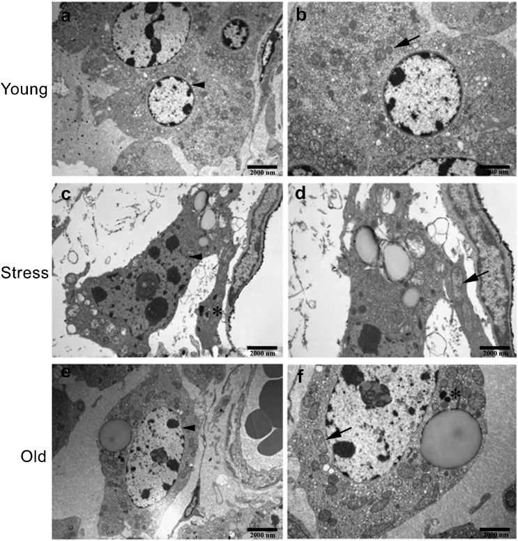
Figure 3.
The ultrastructures of rat Leydig cells were observed using a transmission electron microscope (TEM). (a, b) Images of Leydig cells from young control rats (6-month-old). The Leydig cells were large and satiation and had a normal shape and structure, including abundant smooth endoplasmic reticulum and a prominent rim of heterochromatin beneath the nuclear membrane. (c, d) Images of Leydig cells from stressed rats. (e, f) Images of Leydig cells from old rats (21-month-old). In contrast with Leydig cells from young control rats, aggregation of nuclear chromatin (black arrowhead), dilation of the endoplasmic reticulum, mitochondrial swelling (black arrow), large lipid droplets, nucleolus enlargement and increased lipofuscin accumulation (asterisk) were apparent in Leydig cells from both chronically stressed and aged rats. Scale bars=2000 nm.

