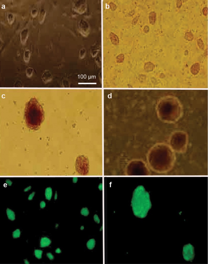
Figure 1.
Morphological characterisation and GFP expression of iPS cells and iPS cell-derived EBs. Phase-contrast microscopy showed the morphological characteristics of iPS cells (a, b) and iPS cell-derived EBs (c, d). Immunofluorescent microscopy revealed GFP expression in iPS cells (e) and iPS cell-derived EBs (f). EB, embryoid body; GFP, green fluorescent protein; iPS, induced pluripotent stem.

