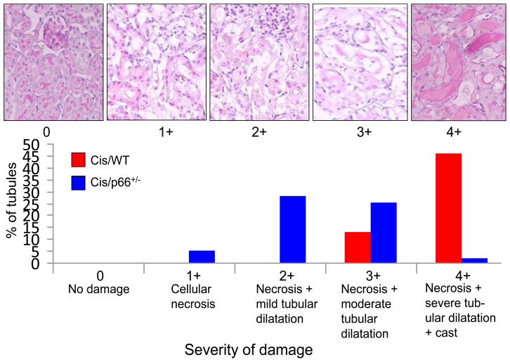
Fig. 1. Cis/p66+/− mice display attenuated tubular cell injury.
A. Representative microphotographs showing different grading of tubular cell injury.
B. Cis/WT and Cis/WT mice (n=6) were graded for their tubular cell injury. Percentages of tubules displaying variable severity of injury are shown displayed massive tubular cell necrosis and dilatation of tubules. Approximately 50% tubules displayed 4+ injury in Cis/WT; whereas, only 1.5% of tubules in Cis/p66+/− displayed 4+ injury.

