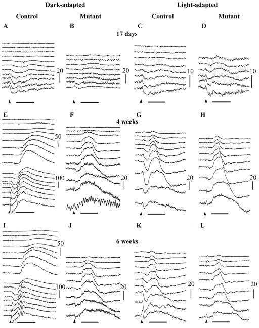
Figure 2.
Representative dark-adapted (A, B, E, F, I & J) and light-adapted (to 30 cd/m2 white light for 10 minutes) (C, D, G, H, K & L) ERG intensity series of a control (A, C, E, G, I & K) and PDE6A mutant (B, D, F, H, J & L) puppy at 17 days (A–D), and 4 (E–H) and 6 (I–L) weeks of age. Key: Arrowheads indicate flash onset. Horizontal bar = 50 mSec, Vertical calibration bars are in microVolts. Flash intensities for dark-adapted were: −3.18, −2.98, −2.79, −2.6, −2.0, −1.6, −1.19, −0.79, −0.39, 0.0, 0.39, 0.85, 1.36, 1.9, 2.38, 2.82 log cdS/m2. Flash intensities for light-adapted were −0.39, −0.22, 0.0, 0.16, 0.39, 0.85, 1.36, 1.9, 2.38, 2.82 log cdS/m2. Note that in the 17 day old puppies (A–D) and the mutant puppies at 4 and 6 weeks of age (F, H, J and L) the tracings from the lower flash intensities are not shown.

