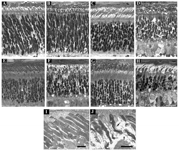
Figure 5.
Progression of outer retinal degeneration in PDE6A mutant puppies. Control (A, C, E, G and I) and mutant (B, D, F, H and J) retinal sections at 2 (A and B), 4 (C and D), 7 (E and F) and 9 (G and H) weeks of age from the central retina (region 4 - Figure 1). I and J; EM at 31 days of age. Sections from normal and mutant are comparable at 2 weeks of age (A and B). By 4 weeks of age there is gross distortion of developing photoreceptor outer segments (D) and dying and pyknotic photoreceptor nuclei are present (arrowheads and arrows respectively). At 7 weeks of age (F) the changes are more severe with further loss of outer segments and thickening of the remaining inner segments, the outer nuclear layer is further thinned. At 9 weeks of age (H) only a few thin outer segments are visible and the outer nuclear layer is thinner and several pyknotic nuclei are present. Note that the remaining photoreceptor inner and outer segments are predominantly of cones and that the inner segments appear abnormally thickened. I and J are EM images of the outer segments in a normal and mutant retina at 31 days of age. The normal regular arrangement of photoreceptor outer segments is lost in the mutant retina with distortion of outer segment discs. A relatively well preserved outer segment (most likely cone) is indicated by an asterix. Arrows indicate remaining outer segments with distorted disc material. The arrowhead indicates part of a thickened inner segment. A–H size bar = 25 μm. I and J size bars = 2 μm.

