
Figure 4.
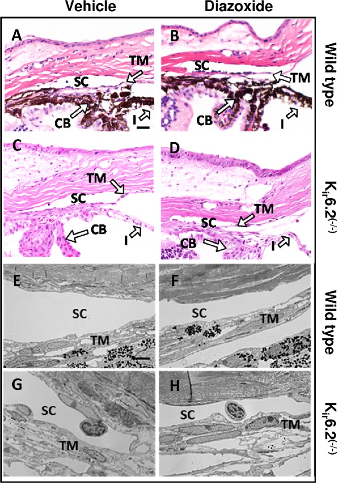
Effect of diazoxide treatment on ocular histology of C57BL/6 wild-type and Kir6.2(−/−) mice. Histologic examination of treated and control eyes in both wild type (A, B, E, F) and Kir6.2(−/−) mice (C, D, G, H) did not show any observable change in cell and tissue appearance within the conventional outflow pathway following diazoxide treatment as evident from H&E staining (A–D) and transmission electron microscopy (E–H). Scale bar: 50 μm for H&E and 5 μm for transmission electron microscopy.