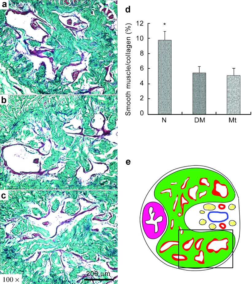
Figure 4.
Masson's trichrome staining. (a–c) Representative images of Masson's trichrome staining of each experimental group: (a) normal group; (b) diabetic group; (c) melatonin treatment group. Original magnification is ×100. Scar bar=200 µm. (d) Results expressed as the ratio between smooth muscle and collagen in corpus cavernosum. (e) The field in the boxed area was used for quantitative analysis. *P<0.05 compared with the diabetic group.

