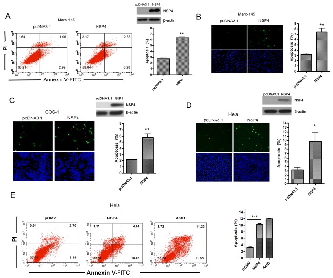
Figure 3. PRRSV nsp4 caused apoptosis in various cell lines.
(A) Flow cytometry analysis of nsp4-induced apoptosis in Marc-145 cells. Cells were transfected with nsp4 expression plasmid or pcDNA3.1 (+) as control. At 48 hours post transfection, cells were collected, stained, and analyzed by flow cytometry. (B, C, and D) In situ TUNEL analysis of nsp4-induced apoptosis in Marc-145, COS-1, and Hela cells. Marc-145 (B), COS-1 (C), and Hela cells (D) were transfected with either pcDNA 3.1 (+) vector or nsp4 expression vector. Forty-eight hours later, cells were fixed, stained with TUNEL reaction mixture, and then detected by fluorescence microscopy. The expression of nsp4 was examined with western blotting using anti-nsp4 serum. (E) Flow cytometry analysis of nsp4-induced apoptosis in Hela cells. Hela cells were transfected with pCMV-Myc control or nsp4-expressing plasmid, or treated with Actinomycin D (ActD) at a concentration of 15 ng/ml as a positive control, respectively. After 48 hours, cells were collected, stained, and analyzed by flow cytometry. Results represent means ± SD of three independent experiments. P<0.01 (**) P<0.001 (***) as determined by student’s t test.

