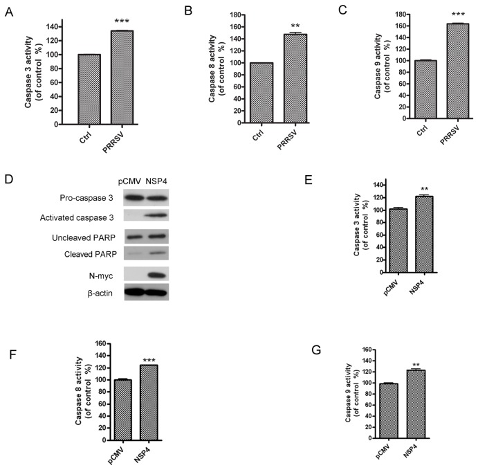
Figure 4. PRRSV-infection and nsp4-transfection activated caspases.
(A, B, and C) PAMs were mock infected or infected with HP–PRRSV (HV strain) at an MOI of 0.5. At 24 hours post infection, the enzymatic activities of caspase-3, -8, and -9 were examined using colorimetric assays. (D) Hela cells were transfected with pCMV-Myc control or nsp4-expressing plasmid. At 48 hours post transfection, activated caspase 3 (17 kDa), cleaved PARP, and nsp4 were analyzed using western blot. (E, F and G) Hela cells were transfected with pCMV-Myc control or nsp4-expressing plasmid. At 48 hours post transfection, the enzymatic activities of caspase-3, -8, and -9 were examined using colorimetric assays. Data represent means ± SD of three independent experiments. P<0.05 (*), P<0.01 (**), P<0.001 (***) as determined by student’s t test.

