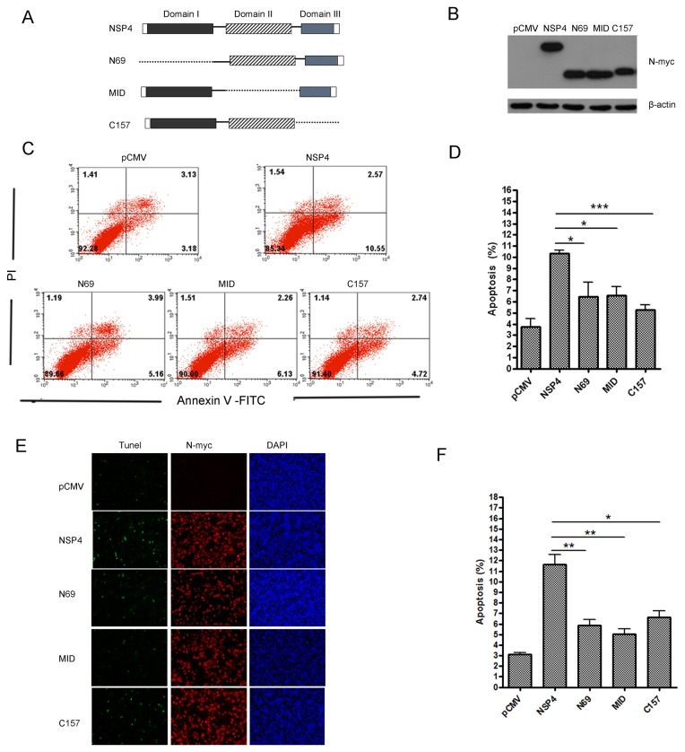
Figure 5. Deletion of either of the three PRRSV nsp4 domains impaired its ability to induce apoptosis.
(A) Schematic diagram represents the PRRSV nsp4 protein deletion mutant constructs. (B) Expression of nsp4, N69, MID, and C157 proteins in Hela cells. Cells were transfected with pCMV-Myc, wild-type nsp4, or deletion mutant plasmids. At 48 hours post transfection, cells were lysed for western blotting to verify proteins expression using anti-myc antibodies. (C) Flow cytometry analysis of wild-type nsp4 and deletion mutants-induced apoptosis in Hela cells at 48 hours post transfection. (D) Percentage of apoptotic cells in panel C. (E) Double-labeling immunofluorescence analysis using TUNEL for apoptosis and indirect immunofluorescence for wild-type and mutant nsp4 proteins in Hela cells at 48 hours post transfection. Apoptosis (green), wild-type or mutant nsp4 (red), and nucleus (blue) were detected by immunofluorescence staining. (F) Percentage of apoptotic cells in panel E. Data represent means ± SD of three independent experiments. P<0.05 (*), P<0.01 (**), P<0.001 (***) as determined by student’s t test.

