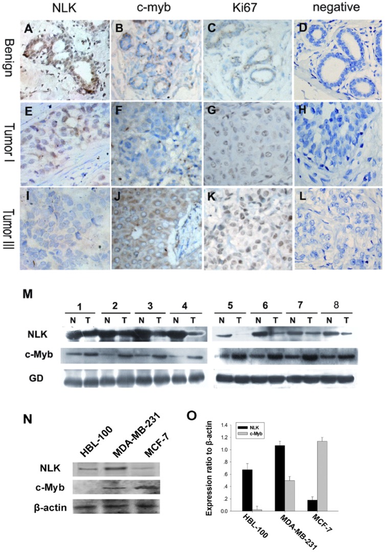
Figure 1. Expression of NLK and c-Myb in human breast cancer.
Paraffin-embedded tissue sections were stained with antibodies for NLK, c-Myb and Ki-67 and then counterstained with hematoxylin. Fig. A–C, E–G High NLK expression was observed in benign breast disease and breast carcinoma specimens (grade I), whereas c-Myb and Ki67 levels were low in the same specimens (SP×400). Fig. I-K High levels of c-Myb and Ki67 were observed in grade III tumor cells. In contrast, NLK expression was low. Fig. 1D, H, and L show negative controls for the benign breast disease and the breast carcinoma specimens. Experimental details are described in the Materials and Methods section. (M) Expression of NLK and c-Myb in eight representative paired samples of breast carcinomas and adjacent normal tissues. (N) Western blot analysis of endogenous NLK and c-Myb in a normal human breast epithelial cell line (HBL-100) and two human breast cancer cell lines (MDA-MB-231 (ER–) and MCF-7 (ER+)). β-actin was used as a loading control. The experiment was repeated at least three times. (O) Quantification indicated that MDA-MB-231 cells displayed the highest levels of NLK and the lowest levels of c-Myb among the two tumor cell lines. In contrast, the lowest NLK and highest c-Myb expression were observed in MCF-7 cells.

