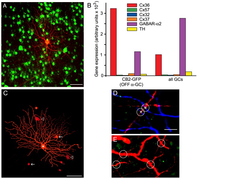
Figure 6. OFF α-GCs Express Cx36 In AC-To-GC, but Not GC-To-GC, Gap Junctions.
(A) Example of the morphology of a single Neurobiotin injected GFP-positive GC (red) in the CB2-GFP retina corresponding to that of an OFF α-GC (41). Scale bar = 50 µm. (B) Relative gene expression of Cx36, Cx57, Cx32, Cx37, GABA receptor α2 subunit (GABAR- α2) and tyrosine hydroxylase (TH) in CB2+ OFF α-GCs. (C) A Neurobiotin-injected OFF α-GC in the WT mouse retina displaying the characteristic tracer coupling to neighboring GCs and ACs. Scale bar = 50 µm. (D) Single optical section (thickness = 0.5 µm) of a confocal image showing the colocalization of Cx36 immunolabeled puncta (green) with dendritic crossings of Neurobiotin-injected OFF α-GC (blue) and tracer coupled ACs (red). Scale bar = 10 µm. (E) No significant colocalization of Cx36 puncta (green) are found at dendritic crossings of Neurobiotin coupled neighboring OFF α-GC dendrites (red).

