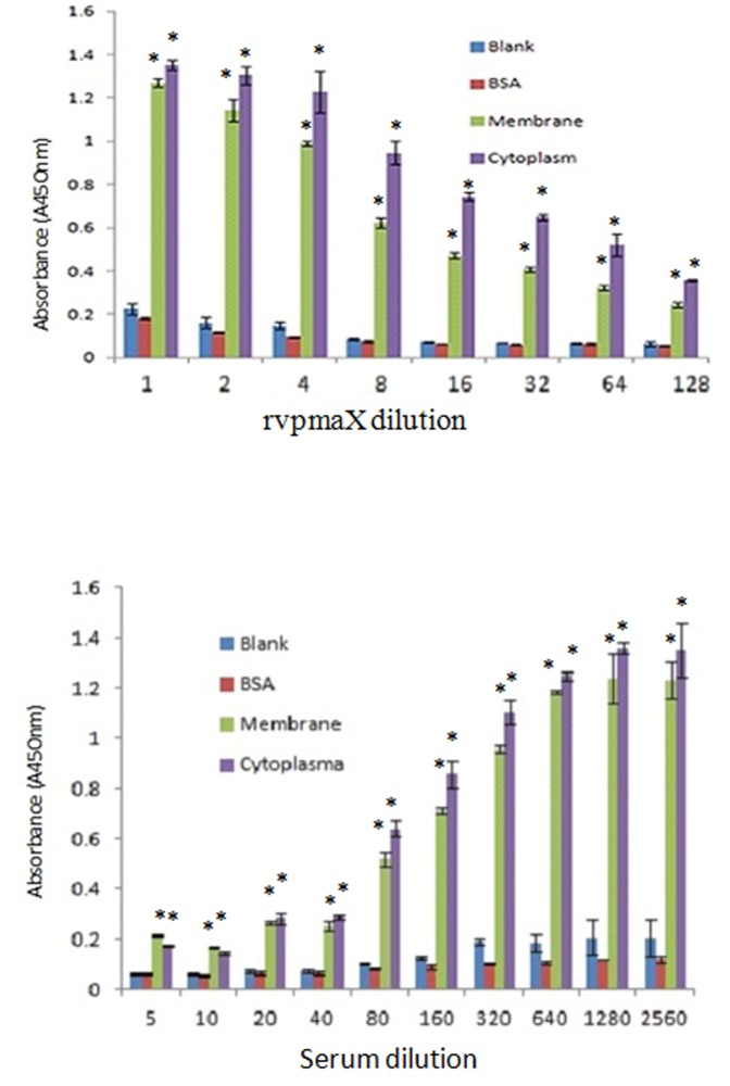
Figure 3. Sandwich ELISA tests of rVpmaX adhesion and adhesion inhibition.
ELISA plates were coated with 1 µg EBL cell membrane or cytoplasmic fraction proteins per well. Plates coated with an equivalent amount of BSA and non-coated plates serve as the negative control and blank control, respectively. (A) 1 µg rVpmaX in 100 µl PBST was serially diluted to 128-fold and incubated with the treated plates. (B) The adhesion of 0.5 µg rVpmaX was inhibited by decreasing concentrations of anti-rVpmaX antiserum (in serial dilutions from 1/5 to 1/2560). *P<0.01, compared with the absorbance of BSA-coated wells in the corresponding group.

