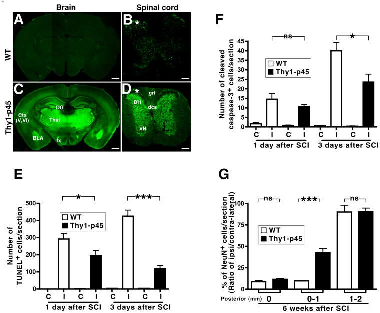
Figure 3. Thy1-p45 transgenic mice display decreased cell death following stab wound injury of the spinal cord.
(A–D) Brain (A, C) and spinal cord (B, D) sections from WT littermates (A, B) and Thy1-p45 transgenic mice (C, D) were immunostained with an anti-p45 antibody. Ctx (V, VI), cortex layers V&VI; BLA, basolateral amygdala; DG, dentate gyrus; Thal, thalamus; fx, fibers of fornix; DH, dorsal horn; VH, ventral horn; grf, gracile fasiculus; dcs, dorsal corticospinal; *, spinal projection fibers from dorsal root ganglia neurons. Scale bar, 1 mm (A, C); 200 µm (B, D). (E–F) WT and Thy1-p45 mice were subjected to stab wound injury in the right side of the spinal cord at the T13 level. One and 3 days after injury, sections were stained for TUNEL+ (E) and activated caspase-3+ (F) cells. The numbers of TUNEL+ and activated caspase-3+ cells were counted in the half spinal cord contralateral (C) and ipsilateral (I) to the stab wound injury. The numbers of TUNEL+ and activated caspase-3+ cells in the ipsilateral side were markedly lower in the Thy1-p45 mice. (G) Six weeks after stab wound injury, the number of NeuN+ cells within 1 mm from injury site was significantly higher in the Thy1-p45 mice compared to their WT littermates, suggesting that p45 expression may decrease neuronal cell death. *P<0.01. ***P<0.0001. ns, not significant. Data are represented as means ± s.e.m.

