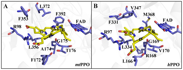
Figure 2. The binding models of protogen in tobacco mtPPO and hPPO.

(A) Side view of protogen surrounded by R98, F172, A174, G175, T176, F353, L356, F392, and FAD in tobacco mtPPO. (B) Side view of protogen surrounded by R97, L166, R168, G169, V170, F331, L334, M368, and FAD in hPPO.
