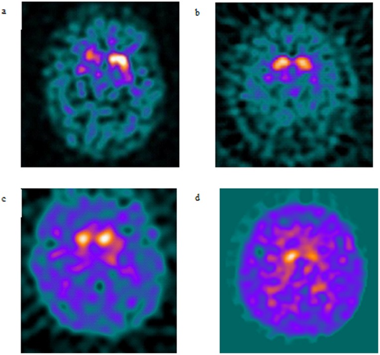
Figure 3. Representative DaTSCAN images.
3a. DaTscan from 50 year old man with GBA mutation, note predominantly right sided tracer loss. 3b. DaTscan from 67 year old woman with PINK1 mutation associated Parkinson's disease, note symmetrical loss of tracer uptake in caudate heads. 3c. DaTscan from 45 year old man with Parkin mutation associated Parkinson's disease and symmetrical loss of tracer uptake. 3d. DaTscan from 32 year old man with LRRK2 mutation, note asymmetrical loss of tracer uptake in caudate heads.

