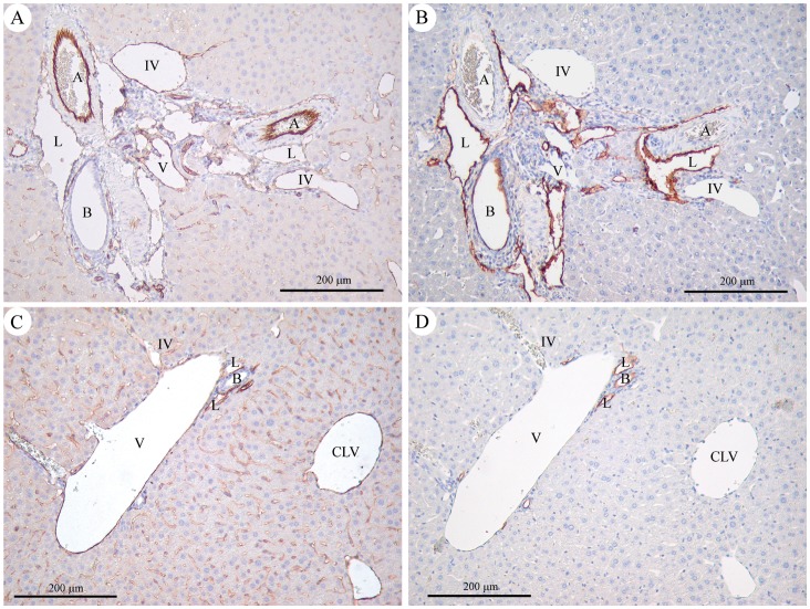
Figure 4. Representative immunohistochemistry findings in a “High Gln” mouse (A, B) and in a “Normal Gln” mouse (C, D).
Original magnification 100x. A and C: CD31 immunostaining highlights endothelial cells. In the “High Gln” mouse (A), the hepatic artery branch is of normal size, similar to that of the interlobular bile duct; the portal vein is small and hypoplastic, and the inlet venules are dilated. In the “Normal Gln” mouse (C), the portal vein is large, with a normal size ratio to the interlobular bile duct (the hepatic artery branch is not seen in this section). The inlet venule is thin. B and D: D2–40 expression confirms the lymphatic nature of the dilated channels at the periphery of the portal tracts in the “High Gln” mouse (B), being selectively reactive in lymphatic endothelial cells, contrary to arterial and venous endothelial cells. Of note, D2–40 (podoplanin) reactivity is also seen in bile duct epithelium. Abbreviations: A = hepatic artery; B = interlobular bile duct; CLV = centrilobular vein; IV = inlet venule; L = lymphatic vessel; V = portal vein.

