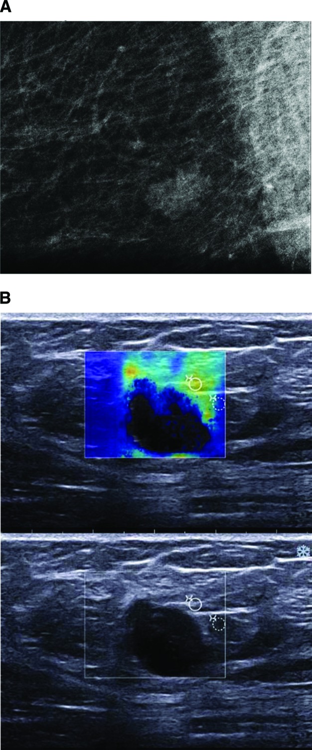
Figure 1.

Imaging studies of a 66-year-old woman with a diagnosis of triple-negative ductal invasive carcinoma. (A): Mammogram shows a microlobulated-shaped mass with mostly circumscribed margins. (B): Ultrasound image shows an oval-shaped mass with microlobulated margins, marked hypogenicity, abrupt interface, and posterior acoustic enhancement. The periphery of the lesion was hard on elastography.
