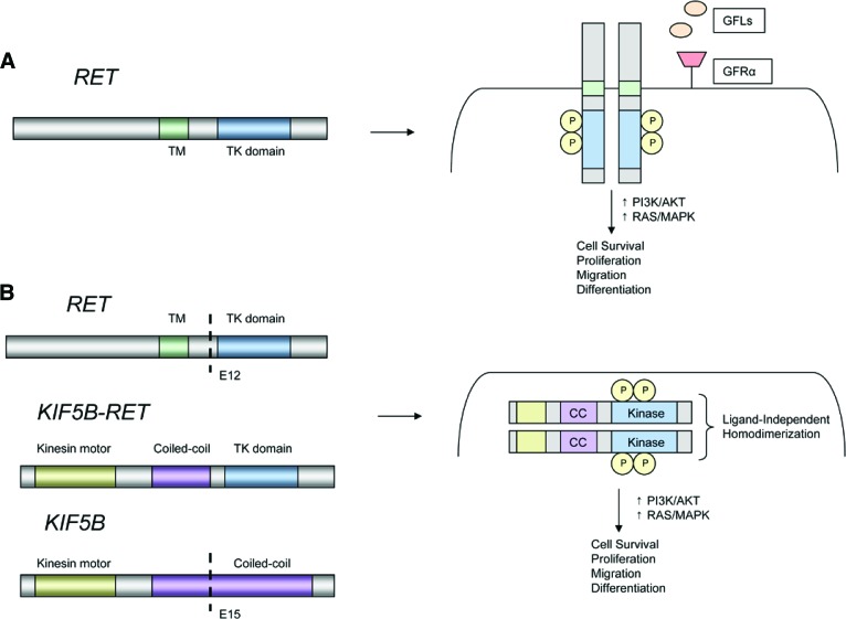
Figure 4.
Models of RET rearrangements. (A): Schematic representation of the RET proto-oncogene (left). RET activation typically involves ligand binding, interactions with a coreceptor, and homodimerization leading to formation of a multiprotein complex (right). (B): Schematic representation of a KIF5B-RET fusion (left). The coiled-coil domain of KIF5B promotes ligand-independent homodimerization of RET, leading to constitutive activation of downstream growth signaling.
Abbreviations: CC, coiled-coil domain; GFL, glial cell line-derived neurotrophic factor family ligand; GFRα, GDNF family receptor α; KIF5B, kinesin family member 5B; P, phosphorylated tyrosine residue; RET, rearranged during transfection; TK, tyrosine kinase; TM, transmembrane.

