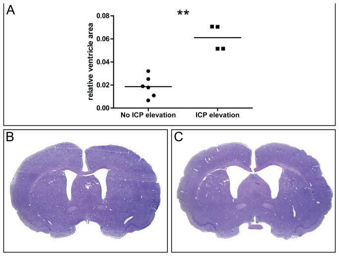
Figure 4. Intracranial pressure and histology.
Relative ventricle area (ventricle area / brain area) (A) in SAH animals with normal ICP (≤ 10 mmHg) or elevated ICP (> 10 mmHg). Representative micrographs of coronal Nissl stained brain sections in a SAH animal with normal ICP (B) and a SAH animal with ICP elevation (C). Mean is shown. ** p<0.01.

