
Fig. 2.
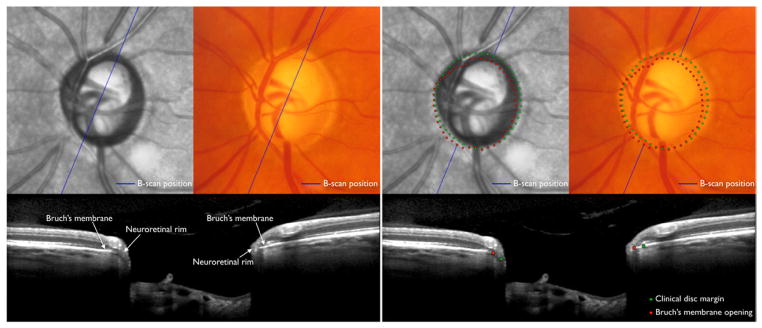
Optic disc margin anatomy with conventional optic disc photography and spectral domain optical coherence tomography (SD-OCT) in the left eye of a patient with glaucoma. Top: Optic disc with infrared illumination (first and third columns) with SD-OCT imaging and photography (second and fourth columns) precisely co-localised. Bottom: Co-localised B-scan images corresponding to the position (blue line) indicated in the fundus images. Clinically visible optic disc margin (green dots; top right) traced in the disc photograph projected to the B-scan (bottom right), and Bruch’s membrane opening identified in the B-scan (red dots; bottom right) projected to the fundus photograph (top right). In this case, there is a clinically invisible overhang of Bruch’s membrane in the superior temporal quadrant resulting in a significant overestimation of the neuroretinal rim width with disc margin based evaluation, while in the inferior nasal quadrant the rim is actually wider than perceived clinically.